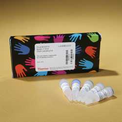
The Thermo Scientific Pierce Biotin 3 End DNA Labeling Kit is for tagging single-stranded DNA primers with biotin for use in non-radioactive electrophoretic mobility shift assays (EMSA) and other nucleic acid detection methods.
The DNA biotinylation procedure uses terminal deoxynucleotidyl transferase (TdT) to catalyze nontemplate-directed nucleotide incorporation onto the 3-OH end of single-stranded DNA. TdT exhibits a substrate preference of single-stranded DNA, but it will label duplex DNA with 3 overhangs and blunt duplexes, albeit with a lower efficiency.
Highlights:
- Non-isotopic labeling eliminates the hassle of hazardous radioactive materials or difficult-to-dispose-of waste
- 1-3 biotinylated ribonucleotides onto the 3 end of DNA strands for less interference with hybridization or sequence-specific binding of proteins
- Biotin-labeled probes are stable for more than one year
- 30-minute labeling procedure is fast and efficient
Product Details:
The Biotin 3 End DNA Labeling Kit has been optimized to incorporate 1-3 biotinylated ribonucleotides (Biotin-11-UTP) onto the 3 end of DNA strands. This labeling strategy has the advantage of localizing the biotin to the 3 end of the probe where it will be less likely to interfere with hybridization or sequence-specific binding of proteins. Biotin-labeled DNA probes can be used to facilitate non-isotopic detection in a variety of applications including electrophoretic mobility shift assays (EMSA), Northern or Southern blots, colony hybridizations orin situhybridizations.
 |
| Sequencing gel analysis confirms efficient biotinylation of oligonucleotides.Ten different oligos (ranging in size from 21-25 nt) were labeled using the Thermo Scientific Biotin 3 End DNA Labeling Kit. The products from the TdT reaction were then radiolabeled using T4 polynucleotide kinase (PNK) and [g-32P]ATP. The PNK reactions were run on a 20% acrylamide/8 M urea/TBE. The position of the starting oligo (no biotin) is denoted by “n.” Incorporation of Biotin-11-UTP by TdT is limited to one or two incorporations per strand (positions “n+1” and “n+2,” respectively). Labeling efficiencies ranged from 72% (EBNA sense strand) to 94% (Oct-1 sense strand). The kit control oligo labeled with 88-94% efficiency. |
 |
| Biotin end-labeled DNA duplexes are effective targets for chemiluminescent gel shift assays.Sense and antisense strands were labeled using the Thermo Scientific Biotin 3 End DNA Labeling Kit and then hybridized for 4 hours at room temperature to form. duplexes containing the binding sites for the indicated transcription factors. Gel shift assays were performed using the Thermo Scientific LightShift Chemiluminescent EMSA Kit using 20 fmol duplex per binding reaction. The source of the transcription factors was a HeLa nuclear extract prepared using Thermo Scientific NE-PER Nuclear and Cytoplasmic Extraction Reagents (Product # 78833) (2 µl or 6-7 µg protein per reaction). In the case of the NF-kB system, nuclear extracts were made from HeLa cells that had been induced with TNFaor cells that were untreated. Competition reactions containing a 200-fold molar excess of unlabeled duplex were performed to illustrate the specificity of the protein:DNA interactions. |
References:
- Cornelussen, R.N.M.,et al. (2001).J. Mol. Cell Cardiol.33, 1447-1454.
- Ho, E. and Ames, B. N., (2002).Proc. Natl. Acad. Sci. USA99(26), 16770–16775.
- Maclean, K. N.,et al.(2004).J Biol. Chem.279(10), 8558–8566.
- Michelson, A.M. and Orkin, S.H. (1982).J. Biol. Chem. 257, 14773-14782.
- Roychoudhury, R. and Wu, R. (1980).Methods Enzymol. 65, 43-62.
- Roychoudhury, R.,et al. (1976).Nucleic Acids Res.3, 863-877.
- Sauzeau, V.et al.(2003)J Biol Chem278(11), 9472–9480.
- Shukla, S. and Gupta, S. (2004).Clin Cancer Res10, 3169-3178.
LightShift Chemiluminescent EMSA Kit
Chemiluminescent Nucleic Acid Detection Module
Related Links:
LightShift Chemiluminescent EMSA FAQ
Modify and label oligonucleotide 5 phosphate groups
Anneal complementary pairs of oligonucleotides |


